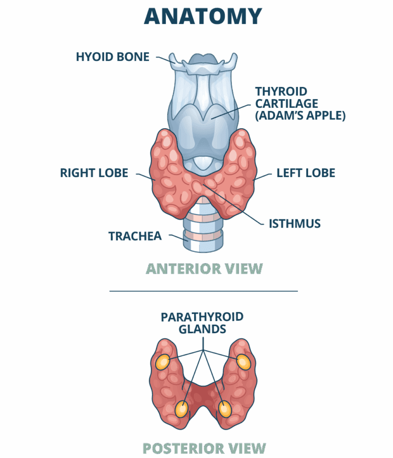
The thyroid gland sits at the front of the neck. It consists of two lobes (left and right lobe) situated on either side of the trachea (windpipe). A thin strip of thyroid tissue (called the isthmus) crosses in front of the trachea and connects the two lobes.
At a microscopic or cellular level the thyroid gland consists of numerous small sacs (called follicles). These sacs are containers for a gel -like protein called colloid. Colloid stores another protein called thyroglobulin (the thyroid hormone building block or pre-cursor). This is the raw material from which the gland produces thyroid hormones.
The thyroid gland sits in contact with or close to many sensitive structures in the neck. Between the trachea and each thyroid lobe lies the recurrent laryngeal nerve which supplies the voice box (vocal cords). The large neck vessels (carotid artery and jugular vein) run along the outer (lateral) edge of each lobe. Underlying the thyroid gland are the tiny parathyroid glands (two on each side). The parathyroid glands are the main regulators of body calcium.